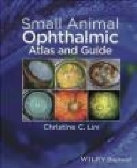
Small Animal Ophthalmic Atlas and Guide
Christine Lim
Small Animal Ophthalmic Atlas and Guide
Christine Lim
- Producent: John Wiley
- Rok produkcji: 2015
- ISBN: 9781118689769
- Ilość stron: 168
- Oprawa: Miękka
Niedostępna
Opis: Small Animal Ophthalmic Atlas and Guide - Christine Lim
Small Animal Ophthalmic Atlas and Guide offers fast access to a picture-matching guide to common ophthalmic conditions and key points related to diagnosing and managing these diseases. The first half of the book presents photographs of ophthalmic abnormalities with brief descriptions, as an aid for diagnosis. The second half of the book is devoted to concise, clinically oriented descriptions of disease processes, diagnostic tests, and treatments for each condition. Small Animal Ophthalmic Atlas and Guide is a useful tool for quickly and accurately formulating a diagnosis, diagnostic strategy, and treatment plan for small animal patients.
Ideally suited for use in the fast-paced practice setting, this text provides both reference images and information for managing the disease in a single text. Small Animal Ophthalmic Atlas and Guide is an easy-to-use aid for small animal general practitioners, veterinary students, and veterinary interns seeking a quick yet complete guide to small animal ophthalmology.
Preface x
List of abbreviations xi
Glossary xii
Section I Atlas 1
1 Orbit 3
Clinical signs associated with orbital neoplasia 3
Clinical signs associated with orbital cellulitis 3
Enophthalmos 4
Brachycephalic ocular syndrome 4
Ventromedial entropion associated with brachycephalic ocular syndrome 4
Clinical signs associated with Horner s syndrome 5
Clinical signs associated with Horner s syndrome 5
Appearance of Horner s syndrome following application of sympathomimetic drugs 5
Clinical signs associated with orbital neoplasia 6
Clinical signs associated with proptosis 6
2 Eyelids 7
Normal appearance of punctum 7
Ectopic cilia 7
Distichiae 8
Distichiae 8
Ectopic cilia and chalazion 8
Lower eyelid entropion due to conformation 9
Facial trichiasis 9
Eyelid agenesis 9
Appearance of entropion after temporary correction using tacking sutures 10
Lower eyelid ectropion 10
Meibomian adenoma 10
Meibomian adenoma 11
Eyelid melanocytoma 11
Chalazion 11
Blepharitis 12
Blepharitis 12
Blepharitis 12
3 Third eyelid nasolacrimal system and precorneal tear film 13
Pathologic changes to the third eyelid associated with pannus 13
Scrolled third eyelid cartilage 13
Prolapsed third eyelid gland ( cherry eye ) 14
Prolapsed third eyelid gland ( cherry eye ) 14
Superficial neoplasia of the third eyelid 14
Neoplasia of the third eyelid gland 15
Positive Jones test appearance of fluorescein in mouth 15
Positive Jones test appearance of fluorescein at nostril 15
4 Conjunctiva 16
Normal appearance of the conjunctiva 16
Conjunctival hyperemia 16
Chemosis and conjunctival hyperemia 17
Chemosis 17
Conjunctival follicles 17
Conjunctival thickening associated with infiltrative neoplasia 18
Superficial conjunctival neoplasia 18
Subconjunctival hemorrhage 18
Conjunctival follicles 19
Superficial conjunctival neoplasia 19
Conjunctival neoplasia 19
5 Cornea 20
Superficial corneal vascularization associated with keratoconjunctivitis sicca 20
Appearance of a dry cornea with concurrent anterior uveitis 20
Typical appearance of superficial corneal vessels 21
Typical appearance of deep corneal vessels 21
Typical appearance of corneal edema 21
Corneal melanosis 22
Typical melanin distribution associated with pigmentary keratitis 22
Typical appearance of corneal fibrosis with concurrent corneal vascularization 22
Corneal white cell infiltrate with concurrent corneal vascularization and edema 23
Corneal deposits associated with corneal dystrophy 23
Corneal deposits associated with corneal dystrophy 23
Corneal mineral deposits 24
Typical changes associated with keratoconjunctivitis sicca 24
Predominantly melanotic corneal changes associated with pannus 24
Predominantly fibrovascular corneal changes associated with pannus 25
Corneal changes associated with pannus mixture of melanosis and fibrovascular changes 25
Superficial corneal ulcer 25
Indolent corneal ulcer with fluorescein stain applied 26
Indolent corneal ulcer prior to fluorescein stain 26
Indolent corneal ulcer after fluorescein stain 26
Deep corneal ulcer 27
Descemetocele after application of fluorescein stain 27
Descemetocele after application of fluorescein stain viewed with cobalt blue light 27
Corneal perforation with iris prolapse 28
Corneal perforation with iris prolapse and hyphema 28
Corneal perforation with large iris prolapse 28
Melting corneal ulcer 29
Perforated melting corneal ulcer 29
Dendritic corneal ulcers 29
Eosinophilic keratoconjunctivitis 30
Eosinophilic keratoconjunctivitis 30
Eosinophilic keratoconjunctivitis 30
Corneal sequestrum; light brown with minimal keratitis 31
Corneal sequestrum; dark brown with moderate keratoconjunctivitis 31
Corneal sequestrum; dark brown with significant keratoconjunctivitis 31
6 Anterior uvea 32
Iris-to-cornea persistent pupillary membranes 32
Iris-to-iris persistent pupillary membranes 32
Posterior synechiae 33
Anterior chamber uveal cysts 33
Anterior chamber uveal cysts 33
Transillumination of posterior chamber uveal cysts 34
Iris atrophy 34
Iris atrophy 34
Severe iris atrophy 35
Focal iris hyperpigmentation 35
Feline diffuse iris melanoma 35
Feline diffuse iris melanoma 36
Canine melanocytoma 36
Canine melanocytoma 36
Canine ciliary body adenoma 37
Clinical signs of uveitis: chemosis episcleral congestion deep corneal vascularization and diffuse corneal edema 37
Aqueous flare 37
Keratic precipitates 38
Hypopyon with concurrent corneal ulceration 38
Hyphema 38
Rubeosis iridis 39
Iris thickening 39
Posterior synechia 39
Iris bombe 40
7 Lens 41
Nuclear sclerosis 41
Nuclear sclerosis 41
Incipient cataract 42
Incipient cataract 42
Incomplete cataract 42
Incomplete cataract 43
Incomplete cataract 43
Complete cataract 43
Complete resorbing cataract 44
Incomplete resorbing cataract 44
Lens subluxation 44
Lens subluxation 45
Anterior lens luxation 45
Posterior lens luxation 45
8 Posterior segment 46
Normal canine fundus 46
Normal canine fundus 46
Normal feline fundus 47
Subalbinotic atapetal canine fundus 47
Subalbinotic feline fundus 47
Tapetal hyperreflectivity and retinal vascular attenuation in a cat 48
Tapetal hyperreflectivity and retinal vascular attenuation in a dog 48
Focal retinal degeneration/chorioretinal scar 48
Focal retinal degeneration/chorioretinal scar 49
Tapetal hyporeflectivity due to hypertensive retinopathy 49
Tapetal hyporeflectivity 49
Tapetal hyporeflectivity 50
Fluid and white cell infiltrate in the nontapetal fundus (chorioretinitis) 50
Focal white cell infiltrate (chorioretinitis) and retinal degeneration in the nontapetal fundus 50
Multifocal retinal degeneration in the nontapetal fundus 51
Hemorrhage in the nontapetal fundus 51
Hemorrhage in the tapetal fundus 51
Serous retinal detachment and retinal hemorrhage due to hypertensive retinopathy 52
External view of serous retinal detachment 52
Serous retinal detachment 52
Rhegmatogenous retinal detachment 53
Optic nerve atrophy/degeneration - mild 53
Optic nerve atrophy/degeneration - severe 53
Optic disc cupping in an atapetal fundus 54
Optic disc cupping 54
Optic neuritis 54
Optic neuritis 55
Asteroid hyalosis 55
Choroidal hypoplasia (collie eye anomaly) 55
Posterior polar coloboma and choroidal hypoplasia (collie eye anomaly) in a subalbinotic fundus 56
9 Glaucoma 57
Typical appearance of acute glaucoma 57
Haab s striae 57
Section II Guide 59
10 Orbit 61
Diseases of the orbit 62
Brachycephalic ocular syndrome 62
Horner s syndrome 63
Orbital cellulitis and abscess 65
Orbital neoplasia 67
Proptosis 68
Further reading 70
References 70
11 Eyelids 71
Diseases of the eyelid 72
Distichiasis 72
Ectopic cilia 73
Trichiasis 73
Eyelid agenesis 75
Entropion 75
Ectropion 77
Eyelid neoplasia 78
Chalazion 79
Blepharitis 80
Eyelid laceration 81
Further reading 82
References 82
12 The third eyelid nasolacrimal system and precorneal tear film 83
Diseases of the third eyelid and lacrimal system 84
Third eyelid gland prolapse ( cherry eye ) 84
Third eyelid neoplasia 85
Nasolacrimal duct obstruction 87
Tear film disorders KCS 88
Qualitative tear film abnormality 88
References 90
13 Conjunctiva 91
Diseases of conjunctiva 91
Canine conjunctivitis 91
Feline conjunctivitis 93
Conjunctival neoplasia 95
Further reading 96
References 97
14 Cornea 98
Corneal diseases 99
Corneal dystrophy 99
Canine keratitis 100
Keratoconjunctivitis sicca (KCS) 101
Pigmentary keratitis 103
Pannus/chronic superficial keratitis 104
Corneal ulceration 105
Simple corneal ulceration 105
Indolent corneal ulceration 106
Deep and perforating corneal ulceration 108
Melting corneal ulceration 110
Feline keratitis nonulcerative and ulcerative 111
Feline eosinophilic keratoconjunctivitis (EK) 112
Corneal sequestrum 113
Further reading 114
References 114
15 Anterior uvea 116
Anterior uveal diseases 116
Persistent pupillary membranes (PPMs) 116
Uveal cysts 117
Iris atrophy 118
Feline diffuse iris melanoma 118
Canine anterior uveal melanocytic neoplasia 119
Iridociliary neoplasia 120
Anterior uveitis 121
Further reading 124
References 124
16 Lens 125
Diseases of the lens 125
Nuclear sclerosis 125
Cataract 126
Lens subluxation and luxation 128
Further reading 129
References 130
17 Posterior segment 131
Diseases of the posterior segment 133
Asteroid hyalosis 133
Collie eye anomaly 133
Progressive retinal atrophy (PRA) 134
Sudden acquired retinal degeneration syndrome (SARDS) 135
Retinal degeneration (excluding PRA and SARDS) 136
Chorioretinitis 137
Retinal detachment 138
Hypertensive retinopathy 140
Optic neuritis 141
Further reading 142
References 142
18 Glaucoma 144
Glaucoma 144
Further reading 148
References 148
Index 149
Szczegóły: Small Animal Ophthalmic Atlas and Guide - Christine Lim
Tytuł: Small Animal Ophthalmic Atlas and Guide
Autor: Christine Lim
Producent: John Wiley
ISBN: 9781118689769
Rok produkcji: 2015
Ilość stron: 168
Oprawa: Miękka
Waga: 0.53 kg


